Friday 28 March 2014

Blood test could predict obesity in children

A simple blood test can measure the levels of epigenetic switches in the PGC1a gene, the gene that governs the body's fat storage.

Obesity is often the product environmental factors: a poor diet and not enough exercise. But people are also -- to varying degrees -- predisposed to obesity.

Recently, researchers at the Universities of Southampton figured out a way to test for obesity. A simple blood test can measure the levels of epigenetic switches in the PGC1a gene -- the gene that governs the body's fat storage.

Epigenetic switches are the subtle changes in gene activity, not sequence, that happen during early development and affect how our genes express themselves in form of physiological traits. These switches take place via chemical changes called DNA methylation, and researchers were recently able to show that higher levels of DNA methylation -- more switching -- predicted a greater likelihood of obesity. Specifically, DNA methylation levels measuring above 10 percent in five-year-olds were associated with a 12 percent increase in body fat nine to ten years later.

It can be difficult to predict when children are very young, which children will put on weight or become obese. It is important to know which children are at risk because help, such as suggestions about their diet, can be offered early and before they start to gain weight. The results of this study provide further evidence that being overweight or obese in childhood is not just due to lifestyle, but may also involve important basic processes that control our genes.

Thursday 27 March 2014

More effort needed to fight hospital infections


A hospital-acquired infection, also known as a HAI, is an infection whose development is favored by a hospital environment, such as one acquired by a patient during a hospital visit or one developing among hospital staff. Such infections include fungal and bacterial infections and are aggravated by the reduced resistance of individual patients.

As a general timeline, infections occurring more than 48 hours after admission are usually considered HAI. These infections are also divided into two classes, endemic or epidemic. Most are endemic, meaning that they are at the level of usual occurrence within the setting. Epidemic infections occur when there is an unusual increase in infection above baseline for a specific infection or organism. HAI occur worldwide, both in the developed and developing world. They are a significant burden to patients and public health. They are a major cause of death and increased morbidity in hospitalized patients. They may cause increased functional disability and emotional stress and may lead to conditions that reduce quality of life. Not only do they affect the general health of patients, but they are also a huge burden financially. The greatest contributors to these costs are the increased stays that patients with these infections require. The increased length of stay varies from 3 days for gynecological procedures to 19.8 days for orthopedic procedures. Other costs include additional drugs, the need for isolation, and the use of additional studies. There are also indirect costs due to loss of work.

Organisms causing hospital infections in India are similar to those around the world, with S. aureus and P. aeruginosa among the most common disease-causing pathogens. Research on hospital infections in India reveals several concerning trends. In Indian ICUs, the rate of vancomycin-resistant enterococcus (VRE), a dangerous hospital infection, is five times the rate in the rest of the world. Rates of methicillin-resistant Staphylococcus aureus in Indian ICUs are also high, with one study finding over 80 per cent of S. aureus samples testing positive for resistance to methicillin and closely related antibiotics.

A large proportion of these hospital infections are easily preventable with increased hospital infection control, including stepping up hygiene practices, such as frequent hand-washing. In India, however, hospitals often do not follow infection control practices, and this leads to the spread of disease. In response to the growing burden of HAIs in India, both the national and international committees on HAI are issuing several key recommendations that aim at reducing the prevalence of HAIs, including increased hand-washing, use of isolation rooms for infected patients, increased availability and uptake of diagnostic tests, reminders to limit catheter use, and use of gloves and gowns. The ministry of health and family welfare task force also recommends that all hospitals create an infection control plan, committee and team. The prevention efforts by them can represent thousands of lives saved, prevented patient harm, and the associated reduction in costs across our country.

Sunday 23 March 2014

Rise in Blood Levels of Heart Muscle Protein an Early Diagnostic Marker for Cardiac Injury

The heart muscle protein cMyBP-C (cardiac myosin binding protein-C) is rapidly released into the bloodstream after cardiac damage and may be a sensitive biomarker for diagnosing the onset of myocardial infarction (MI).

Myosin-binding protein C is a myosin-associated protein found in the cross-bridge-bearing zone (C region) of A bands in striated muscle. It is found at regularly spaced intervals and is hypothesized to act like a "barrel hoop" and hold the thick filament together. cMyBP-C, the cardiac isoforms of the protein, is expressed exclusively in heart muscle.

Detection of elevated plasma cardiac troponin (cTn) levels is the "gold standard" for early discovery of MI. However, troponin levels peak only four to six hours after heart attack and lack the sensitivity required to detect the onset of MI at its earliest stages.

Investigators at the Loyola University School of Medicine (Chicago, IL, USA) sought to evaluate the usefulness of cMYBP-C as an ultra-early biomarker of MI. To this end, they studied the release kinetics of cMyBP-C in a porcine model of MI and in two human cohorts.

They measured cMyBP-C levels in serum and plasma samples from MI pigs and patients serially from 30 minutes to 14 days after coronary damage using a custom immunoassay based on electrochemiluminescence. The anti-cMyBP-C antibodies used in this study were specific to cMyBP-C and were generated against the C0 domain of cMyBP-C, which is exclusively present in the cardiac isoform and does not cross react with skeletal MyBP-C isoforms. The assay was comparable to a previously described ELISA and used the same capture and detection antibodies. The chemiluminescence immunoassay had improved sensitivity, compared to ELISA.

Results revealed that in the pig model system cMyBP-C plasma levels were increased from baseline (around 76 nanograms per liter) to about 767 nanograms per liter three hours after onset of cardiac damage and then peaked at about 2,418 nanograms per liter after six hours. Plasma troponin and myosin light chain levels were all increased after six hours. In a cohort of 12 patients with hypertrophic obstructive cardiomyopathy, cMyBP-C was significantly increased from baseline about 49 nanograms per liter in a time-dependent manner, peaking at about 560 nanograms per liter after four hours. In a second cohort of 176 patients with non-ST segment elevation, cMyBP-C serum levels were significantly higher (about 7,615 nanograms per liter) than those in a control cohort of 153 normal individuals (about 416 nanograms per liter).

“This is a potential ultra-early biomarker that could confirm whether a patient has had a heart attack, leading to faster and more effective treatment,” said senior author Dr. Sakthivel Sadayappan, assistant professor of cell and molecular physiology at Loyola University School of Medicine. “These findings suggest that cMyBP-C has potential as an ultra-early biomarker for the diagnosis of [heart attack], but this still needs to be validated using a large cohort study. A cMyBP-C blood test might lead to an earlier diagnosis in patients who present at the emergency department shortly after coronary artery blockage. However, a systemic prospective investigation is required to establish such data for clinical use.”

The study was published in the February 15, 2014, issue of the American Journal of Physiology – Heart and Circulatory Physiology.


Credits: Lab Medica

Saturday 22 March 2014

Blood Test Identifies Seniors At-Risk for Cognitive Impairment or Alzheimer's Years in Advance of Onset

A team of researchers has discovered biomarkers and validated the first blood test that accurately predicted whether an asymptomatic person will develop amnestic mild cognitive impairment or Alzheimer’s disease within 3 years before onset of clinical symptoms, heralding the potential for developing treatment strategies to prevent, reverse, or slow disease progression.

In a collaborative study between several institutions, mainly Georgetown University Medical Center (GUMC; Washington DC, USA) and University of Rochester School of Medicine (URSM; Rochester, NY, USA), a blood-based biomarker panel has been discovered that may identify preclinical Alzheimer's disease. In the report, published in the journal Nature Medicine, March 9, 2014, online ahead of print, Mapstone et al. describe their lipidomic approach examining cognitively normal senior adults. They discovered and validated a set of lipids from peripheral blood plasma that predicted with over 90% accuracy the phenoconversion within 2-3 years from cognitively normal to either amnestic mild cognitive impairment (aMCI) or on to Alzheimer's disease (AD).

The study has yielded the first blood test that accurately identified individuals at risk for developing AD. The discovery could be a key to unlocking a new generation of treatments that seek to head off the disease before neurological damage becomes irreversible. “Our novel blood test offers the potential to identify people at risk for progressive cognitive decline and can change how patients and their families, and treating physicians plan for and manage the disorder,” said corresponding author Howard J. Federoff, MD, PhD, professor of neurology at the GUMC. Some of the same experimental treatments that have thus far failed may prove to be successful if they are given to high-risk patients much sooner.

Biomarkers of preclinical disease with high sensitivity and specificity are critical. Current biomarkers and methods used in attempt to detect early disease are very limited. For widespread use and large-scale screening, blood-based biomarker screening would be more attractive and may also be more effective.

Wednesday 19 March 2014

LDL is “good,” HDL is “bad,” and triglycerides aren’t really cholesterol at all!


Cholesterol doesn’t dissolve well in the blood. It needs to attach itself to fatty protein to circulate through the body. When we talk about the specific types of cholesterol, we’re actually talking about the different types of proteins that carry the cholesterol molecules through the bloodstream. These types are:

LDL (low-density lipoprotein) cholesterol: LDL is one of the two most important fatty proteins (HDL is the other). LDL cholesterol is usually referred to as “bad” cholesterol because it deposits its cholesterol on the walls of arteries. LDL is also the type of cholesterol that becomes oxidized and damages the lining of your arteries, setting the stage for mineral and fat deposits.

HDL (high-density lipoprotein) cholesterol: Unlike LDL, HDL hangs on tightly to the cholesterol it carries and won’t let it get loose to attach to arterial walls. In some cases, it may even snatch up additional cholesterol already stuck to a wall, reducing the size of a plaque or buildup. HDL keeps cholesterol in solution and moves it safely throughout the body. For these reasons, HDL cholesterol is considered to be “good” cholesterol.

Beta-VLDL (very low-density lipoprotein) cholesterol: Beta-VLDL deposits cholesterol in the small arteries supplying the heart. It receives very little attention because it makes up only a small portion of the total blood cholesterol.

Though they’re not a form of cholesterol, triglycerides are a type of blood lipid that is often lumped together with cholesterol because many of the strategies for reducing triglyceride levels are the same as those for lowering cholesterol. High triglyceride levels are indicative of excess sugar in the diet, and often too much fat. The excess sugar is combined with the fat and typically stored around the gut (the “spare tire”) and around muscles (flabby arms).

Tuesday 18 March 2014

Are your Kidneys OK?

Kidney problems do not develop overnight. The kidney function naturally starts declining after a person cross 30s. And whether you will develop a kidney problem later in life is largely dependent on your lifestyle habits and diet. In most cases, kidney problems are caused as a result of a primary condition like diabetes, hypertension or high cholesterol levels. Here are some kidney function tests that can determine whether you are at a risk of kidney disease.

Blood pressure: Kidneys perform the function of filtering blood under pressure to eliminate waste products through the urine. If your blood pressure is higher than normal it will increase the load on your kidneys and may lead to kidney disease. Alternatively, a person is suffering from kidney disease is also likely to have high blood pressure.

Blood creatinine test: Creatinine is a waste product that is produced by your muscles. It enters the bloodstream and is filtered by the kidneys to get excreted through the urine. A blood creatinine test will measure the amount of creatinine in your blood. A higher level (greater than 0.8-1.2 mg/dl in men and 0.6-0.9 mg/dl in women) may indicate that the kidneys are not functioning properly.

Blood urea test: Urea is a waste product filtered by the kidneys and excreted through the urine. It is produced by breakdown of proteins. A high level of urea in the blood (greater than 20-40 mg/dL) may indicate altered kidneys function.

Urinalysis: It is one of the recommended kidney function tests by the National Kidney Foundation (NKF). It is used to detect the presence of protein and blood cells (RBCs and WBCs) in the urine. Increased levels of both protein and blood cells indicate early signs of kidney disease.

Urea clearance test: This test is based on a comparison between the amount of urea in the blood and the amount of urea in urine. If the urea clearance is lower than the normal range (64-99 ml/min), it may indicate that the kidneys are not working properly.

Creatinine clearance test: A creatinine clearance test also compares the level of creatinine in blood with creatinine in urine to determine the rate at which the kidneys are filtering creatinine. Altered creatinine clearance range (normal range: 85-125 ml/min in men and 75-115 ml/min in women) indicates altered kidney function.

eGFR (estimated glomerular filtration rate):
Glomerular filtration rate is the rate at which your kidneys filter waste products from the blood. eGFR is a value that is derived from a formula based on the amount of blood creatinine, age and gender of the person. According to the NKF, eGFR is the best estimate of determining accurate kidney function. Normal eGFR results range from 90-120 mL/min/1.73 m2.

Renal ultrasound test: Renal ultrasound is an imaging test that uses high frequency ultrasonic waves to check the shape and size of the kidneys. It can detect the flow of blood to the kidneys as well as the presence of tumors, cysts and infection around the kidneys. It also helps to detect presence of kidney stones and external damage or injury to the kidneys

Because the symptoms of kidney diseases are not evident unless the kidneys are deteriorated to a great extent , one should regularly screen your kidney function (especially if suffering from heart disease and diabetes).

Monday 17 March 2014

Patients with even slightly high blood pressure 'at increased risk of stroke'


It is well known that high blood pressure increases the risk of stroke - the fourth leading cause of death worldwide. The threshold for high blood pressure diagnosis, or hypertension, is 140/90 mmHg. But new research suggests that even people with blood pressure lower than this - but higher than normal - have an increased stroke risk.

For the study, the investigators analyzed all existing research that looked at the risk of stroke in people with prehypertension. This is when blood pressure is higher than normal (120/80 mmHg) but below the high blood pressure diagnosis threshold. The analysis included 19 studies involving more than 760,000 participants. All subjects were followed for between 4 and 36 years, and 25-54% of them had prehypertension.

The researchers found that individuals with prehypertension were 66% more likely to have a stroke than those who had normal blood pressure, and almost 20% of strokes that occurred in the study population were in those with prehypertension. These results remained even after the team adjusted for factors that may increase stroke risk, including smoking, diabetes and high cholesterol.

As part of the study, the researchers divided individuals with prehypertension into two groups. Participants in the "high" group had blood pressure over 130/85 mmHg, while those in the "low" group had blood pressure under this but above normal. This analysis revealed that participants in the high group were 95% more likely to have a stroke than those with normal blood pressure, while those in the low group were 44% more likely to have a stroke.

These findings, if confirmed, have important takeaways for the public. Considering the high proportion of the population who have higher than normal blood pressure, successful treatment of this condition could prevent many strokes and make a major difference in public health.

Friday 14 March 2014

Study results could lead to blood test that may detect concussion

New research has found that a protein indicating the breakdown of white matter in the brain can be detected following a hard blow to the head, raising hopes that a blood test could soon detect a concussion, predict how long symptoms will linger, and guide decisions about an athlete's return to play. In their bid to find a blood "biomarker" for concussion, researchers went to a sport with plenty of blood and plenty of concussions: hockey.

Elevated levels of the protein known as total-tau, or T-tau, have long been seen as a marker for brain injury, and are present in a wide range of neurodegenerative disorders, including Alzheimer's disease.

The latest study, published in the journal JAMA Neurology, finds that T-tau levels spiked in the blood in the hour after a hockey player suffered a traumatic brain injury, and then again about 36 hours later. And while they declined slightly in between and after 36 hours, blood samples of players with concussions showed elevated levels of T-tau six days after their injuries, compared with blood samples taken in the pre-season. The higher a player's T-tau level was in the first hour after injury, the longer it took for his concussion symptoms to fade away, researchers found.

While researchers suspected T-tau might be key to diagnosing concussions, detecting elevated T-tau levels is more easily said than done: With varying degrees of accuracy, it can be done by drawing cerebro-spinal fluid from the brain's fluid-filled ventricles, or from spinal fluid. Jabbing a plunger into the brain or the spine is riskier and more expensive than drawing blood and testing it, but no existing blood assays were capable of detecting elevated T-tau levels.

Then, in 2012, two of the study's co-authors, from Quanterix Corp. in Massachusetts, pilot-tested a highly sensitive new immunoassay. They found it capable of detecting heightened T-tau levels in the blood of patients who had hypoxic injuries to the brain after a heart attack. The same assay was used to compare players' blood levels of T-tau from the pre-season to those immediately after a blow to the head.

The same researchers found two other potential biomarkers of little use in distinguishing those who sustained concussion from those who did not.

Iron deficiency increases stroke risk by making blood sticky

More than 15 million people worldwide suffer a stroke every year, resulting in almost 6 million deaths. Now, new research from Imperial College London in the UK finds that iron deficiency could increase a person's risk of stroke by making the blood sticky.

The research team notes that previous research has shown that iron deficiency could be a risk factor for ischemic stroke - when small blood clots interrupt blood flow to the brain - in adults and children.

To investigate why this is the case, the researchers analyzed the iron levels of 497 patients with hereditary hemorrhagic telangiectasia (HHT) - a rare disease than can lead to enlarged blood vessels in the lungs.

The research team explains that healthy blood vessels usually filter out small blood clots before the blood travels to the arteries. But in HHT, the blood vessels can allow small blood clots to make their way to the brain.

The investigators found that patients with moderately low iron levels (6 micromoles per liter) had double the risk of stroke, compared with patients with iron levels deemed middle of the normal range (7-27 micromoles per liter). Further investigation revealed that iron deficiency increases the stickiness of platelets - small blood cells. This prompts platelets to stick together, causing clotting.

Since platelets in the blood stick together more if you are short of iron, this may explain why being short of iron can lead to strokes, though much more research will be needed to prove this link. The research team plans to investigate whether treating iron deficiency in high-risk patients could reduce their risk of stroke, and specifically, whether this would cause platelets in the blood to become less sticky.

Tuesday 11 March 2014

“Over diagnosis” may be responsible for rise in thyroid cancer incidence!

According to a new study published increased incidence of thyroid cancer may not be because of an increase in the disease, but an increase in diagnosis. Several types of cancer can affect the thyroid - a gland in the neck that produces hormones. Factors that make people more at risk of getting thyroid cancer include- being between the ages of 25 and 65, being female, being Asian, having a history of thyroid disease in the family and having previously had radiation treatments to the head or neck.

To diagnose thyroid cancer, doctors use a combination of physical exam, blood tests, imaging tests and a biopsy. Since 1975, the incidence of thyroid cancer has nearly tripled, from 4.9 to 14.3 in every 100,000 people.

To assess why there has been an increase in diagnoses of thyroid cancer, the authors behind the new study analyzed the medical records of patients between 1975 and 2009 in Atlanta. Despite the increase in diagnosis, the researchers did not find an increase in the rates of death from thyroid cancer. About 0.5 per 100,000 people die from this cancer, which has remained stable since 1975.

Because of this, the researchers suspected that "over diagnosis" may be responsible for rise in thyroid cancer incidence. Over diagnosis is when a patient is diagnosed with having a condition that has no symptoms and may never cause them any harm. The researchers found that increased detection of small papillary cancers - a less aggressive form of thyroid cancer - is responsible for the increase in thyroid cancer incidence.

As a response to this over diagnosis, the researchers make several suggestions. They think that some of the small papillary cancers may benefit from not being labeled as cancer, and instead of treating the small papillary cancers, monitoring them through active surveillance instead.

Doctors should explain to patients that many of these small cancers will never grow or cause them any harm, the researchers say. The researchers do concede, though, that it is not possible to know in advance which of these diagnosed cancers will continue to be small and not cause symptoms and which will grow to be a threat to the patient's health.

High blood pressure reading in kids linked to triple risk for condition as adults

Children with one or more high blood pressure readings were about three times more likely to develop the condition as adults, in a study presented at the American Heart Association High Blood Pressure Research Scientific Sessions 2013.

After accounting for age, gender and weight, researchers found a direct link between high blood pressure readings during childhood and high blood pressure in adulthood. The rate of high blood pressure during adulthood was 8.6 percent among those who didn’t have high readings as children. The rate rose to 18 percent among those who had at least one high reading during childhood. The rate jumped to 35 percent among those who had two or more high readings during childhood.

The findings suggest even occasional spikes in blood pressure at any age could signal problems later in life and should not be dismissed.

In 1986, researchers began following 1,117 adolescent children who lived in Indianapolis. Blood pressure readings were taken by a school nurse or during a doctor’s office visit, and the children were followed for 27 years. Among the study participants, 119 were diagnosed with high blood pressure as adults. Fifty-nine percent of the adults diagnosed with high blood pressure had been overweight or obese as children. Childhood obesity is a risk factor for high blood pressure and heart disease in adulthood.

This study highlights the need for pediatricians to regularly check blood pressure and weight. An occasional increase in blood pressure does not justify treatment, but it does justify following these children more carefully.

Friday 7 March 2014

Liver related disorders: Why screening is essential

An average of 35-40 people die every month from liver related disorders in our country. Contrary to the popular belief, even non-alcoholics can acquire deadly liver diseases. There are usually no early symptoms. Doctors say that in more than 90% cases, patients don’t realize that they have a liver disease until it’s late.

There is a major misconception about liver disease- It only affects alcoholics and drug users. Liver disease is not a disease specific to alcohol drinkers. There are more than 100 causes of liver disease. Liver problems can strike anyone, from infants to seniors. Liver disease can be hereditary, caused by toxic substances, viruses and bacteria or an unhealthy lifestyle.

In the early stage of liver disease, liver may become inflamed. However, unlike most other parts of our body that become hot and painful when inflamed, an inflamed liver may cause no discomfort at all. Liver inflammation can slowly worsen, causing scarring or fibrosis to appear. As fibrosis worsens, cirrhosis develops and the liver becomes seriously scarred, hardened and unable to function properly. By the time we experience symptoms such as jaundice, our liver would have been severely damaged.

Major diseases of the liver include autoimmune hepatitis, cirrhosis, cystic disease of the liver, fatty liver disease, gallstones, liver cancer, liver disease in pregnancy, neonatal hepatitis, toxic hepatitis, viral hepatitis A, B& C. Probability of majority of these diseases can be simply screened by performing a full panel liver function test, which indicates how healthy your liver is.

Doctors recommend comprehensive liver checkup once in every 6 months for a normal healthy individual. Liver tests are highly recommended in those with a history of liver disorder in family, people with some sort of life style diseases like diabetes & cholesterol, who lead sedentary life style, who are obese, those who consumes alcohol, under stress, under long term medication etc. Always remember- It’s an organ you could easily trash if you don’t take good care of it, and once you trash it, it’s gone.

Thursday 6 March 2014

The color of your urine says about health

Urine and urinalysis have, for hundreds of years, been one of the ways physicians have looked at health. From a historical view, urinalysis was one of the original windows into what's happening in the body. That's because many of the substances circulating in our body, including bacteria, yeast, excess protein and sugar, eventually make their way into the urine. Urine is an important part of the body's disposal process. Its job is to remove the extra water and water-soluble wastes the kidneys filter from the blood. It’s important to note that the colour of urine says something abou your health. Before you flush, here are a few urine changes to look out for, and what they might be saying about your health:

Clear: Clear urine means that you are well hydrated possibly due to intake of large amounts of water. It's okey to see that.

Orange: When you eat certain foods like carrots or take certain medications. The outcome may tend to be orange urine. It may indicate that there is an excess of Vitamin C in your system. Which is a good thing, however you could cut down on such food stuffs.

Brown: Brown urine can be caused by certain foods, like fava beans or even laxatives. This colour could be a sign of a serious condition. Liver disease, melanoma cancer and hepatitis can all cause urine to have a brown tinge. You may want to report this to a doctor.

Green: A urinary tract infection (UTI) or certain drugs can cause your urine to look a bit green. Few drugs can actually do that. If your slightly green urine is also accompanied by discomfort or burning while you urinate, there is a problem. So basically, when you see green, please see your doctor.

Blue: I was also shocked that there is blue. Urine with a slight blue tint is an indicator of high calcium levels. You can cut down on calcium food( milk or yoghurt,cheese, almonds) or talk to a doctor about your diet to fix this problem. It could also be caused by certain bacterial infections which your doctor will let you know.

Red: Red urine can be caused by many things. Large amounts of red food dye or naturally red foods can change the color in the toilet. However, red urine could probably be an indication presence of blood-Resulting from internal injuries or bladder infection. You may want to visit your doctor as soon as possible.

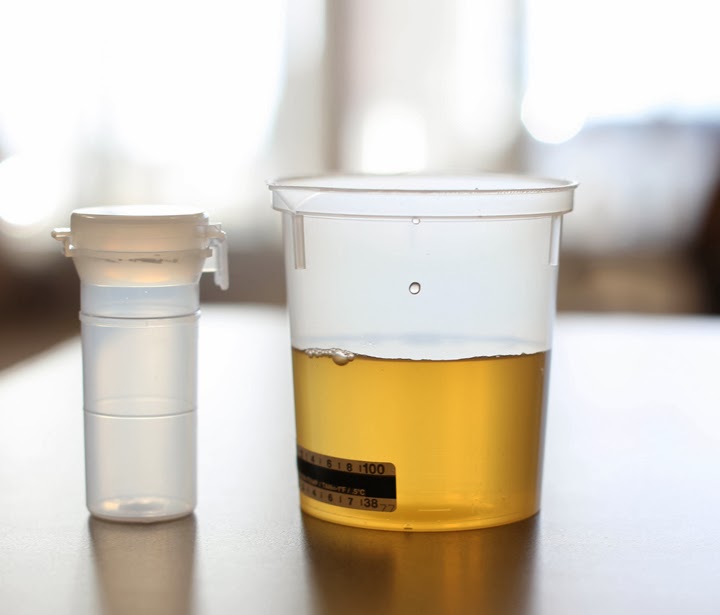
Dark yellow: This is an indication of a few things happening in your system. Most commonly it tells you that you not drinking enough water. Curb this by increasing the water intake. It may also indicate jaundice or liver problems. Someone with jaundice has yellow on the whites of their eyes.

Take good care of your bladder, and it will thank you by helping you urinate regularly. To avoid having to make too many bathroom visits, stay hydrated, but not overhydrated. Drink whenever you're thirsty, but don't feel as though you have to adhere to the eight-glasses-a-day recommendation (unless you have kidney or bladder stones, in which case you'll need to increase your fluid intake). If you're getting up during the night to use the bathroom, stop drinking three to four hours before bedtime. Limit caffeine, which can irritate the lining of the bladder. Also watch your intake of alcohol, which can have similar effects.

Tuesday 4 March 2014

Scientists revive ‘biggest ever’ virus locked in Siberian ice for 30,000 years

A Stone Age virus lying dormant for at least 30,000 years has been ‘successfully’ revived by scientists in Siberia. The newly thawed virus is the biggest one ever found. At 1.5 micrometers long, it is comparable in size to a small bacterium. Tests show that it attacks amoebas, which are single-celled organisms, but does not infect humans or other animals.

The pithovirus sibericum was unleashed from a 100ft deep layer of the Siberian permafrost. Though this virus poses no danger to humans or animals, the discovery has raised fears other more deadly pathogens could be released from the frozen ground. If it is true that these viruses survive in the same way those amoeba viruses survive, then smallpox is not eradicated from the planet - only the surface, Dr Chantal Abergel, a co-author of the report published in the Proceedings of the National Academy of Sciences said. "By going deeper we may reactivate the possibility that smallpox could become again a disease of humans in modern times."

However, it is not yet clear whether all viruses could become active again after being frozen for thousands or even millions of years.

Monday 3 March 2014

Rising Diabetic Population Drives Demand for Hemoglobin A1c Monitoring

Hemoglobin A1c (HbA1c) monitoring is one of the rapidly growing markets in the diagnostics industry. Hemoglobin A1c test for diabetes monitoring is attracting the attention of healthcare providers and patients alike given its numerous advantages, including the ability to perform tests at any given time in a day without the need for overnight fasting, which is a pre-requisite for traditional oral glucose tolerance test. HbA1c test results also provide physicians with information needed to develop intervention strategies and treatment plans to prevent diabetes-related complications.

With diabetes assuming epidemic proportions, there exists strong potential for diabetes monitoring devices including Hemoglobin A1c blood test kits. Aging population, changing lifestyles, and rising obesity levels are contributing to increased number of diabetics worldwide, particularly in countries such as China, India, the United States, Brazil, Russia, Mexico and Indonesia. The need for effective monitoring of diabetes is driving growth in the HbA1c monitoring market. Increasing patient awareness about the advantages of regular monitoring of blood glucose levels in the prevention and control of diabetes is further expected to enhance the average number of tests being performed, thus translating into favorable opportunities for the HbA1c monitoring market.

As stated by the new market research report on Hemoglobin A1c Monitoring, the United States represents the largest market worldwide. Asia-Pacific is forecast to emerge as the fastest growing regional market. With the number of diabetics continuing to increase owing to rapid urbanization, poor dietary habits, and sedentary lifestyles, developing countries offer favorable opportunities for growth. Growing awareness levels coupled with the test’s ability to identify individuals at high risk of developing diabetes favors market growth. Though high cost remains a concern, the need to conduct HbA1c tests on a quarterly basis as opposed to monthly blood sugar level test will likely act in favor of HbA1c testing and systems.

Segment wise, Laboratory-based HbA1c monitoring represents the largest segment in the global HbA1c monitoring market. Laboratories make use of HbA1c analyzers to provide accurate measurement of HbA1c levels in patients. Though laboratory testing dominates the market, the segment faces stiff challenge from the growing popularity of POC testing. Point-of-Care (POC)-based HbA1c monitoring is poised to grow at fastest with a CAGR of 12% over the analysis period, driven by rising incidence of diabetes, convenience benefits, continuous improvements in test reliability and increasing adoption by physicians.